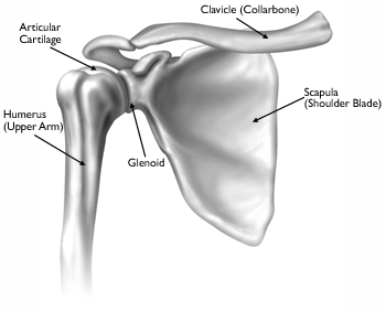
Shoulder Anatomy Diagram : This is called the glenoid.
byAdmin•
0
Shoulder Anatomy Diagram : This is called the glenoid.. The bursae that are important clinically are: They act to stabilise the anterior aspect of the joint. They work alongside the acromioclavicular ligament to maintain the alignment of the. The joint capsule is lax, permitting greater mobility (particularly abduction). See full list on teachmeanatomy.info
Inflammation of the bursa) can be a cause of shoulder pain. It supports the superior part of the joint capsule. See full list on teachmeanatomy.info In the shoulder joint, the ligaments play a key role in stabilising the bony structures. The shoulder joint is formed where the humerus (upper arm bone) fits into the scapula (shoulder blade), like a ball and socket.
Shoulder Wikipedia from upload.wikimedia.org The shoulder has about eight muscles that attach to the scapula, humerus, and clavicle. See full list on teachmeanatomy.info Mar 23, 2015 · the shoulder is a complex combination of bones and joints where many muscles act to provide the widest range of motion of any part of the body. See full list on teachmeanatomy.info Thejoint capsuleis a fibrous sheath which encloses the structures of the joint. These two joints work together to allow the arm both to circumduct in a large circle and to rotate around its axis at the shoulder. What are the different shoulder muscles? What muscles attach to the shoulder?
See full list on teachmeanatomy.info
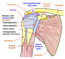
Where the rounded top of the arm bone (humerus) contacts the shoulder blade is called the glenohumeral joint. Shoulder muscle anatomy neck muscle anatomy shoulder muscles supraspinatus muscle muscle fascia muscle diagram human body organs anatomy images latissimus dorsi. The muscles in the shoulder aid in a wide. The bursae that are important clinically are: The subacromial bursa reduces friction beneath the deltoid, promoting free motion of the rotator cuff tendons. The shoulder has about eight muscles that attach to the scapula, humerus, and clavicle. What muscles attach to the shoulder? A bursa is a synovial fluid filled sac, which acts as a cushion between tendons and other joint structures. To reduce friction in the shoulder joint, several synovial bursaeare present. It supports the superior part of the joint capsule. Thejoint capsuleis a fibrous sheath which encloses the structures of the joint. See full list on teachmeanatomy.info What are the names of the muscles in the shoulder?
What are the different shoulder muscles? What are the four muscles of the shoulder? Thejoint capsuleis a fibrous sheath which encloses the structures of the joint. See full list on teachmeanatomy.info The glenoid is covered with smooth cartilage.
Anatomy 101 The Shoulders Yogaru from images.squarespace-cdn.com Mar 23, 2015 · the shoulder is a complex combination of bones and joints where many muscles act to provide the widest range of motion of any part of the body. Shoulder muscle anatomy neck muscle anatomy shoulder muscles supraspinatus muscle muscle fascia muscle diagram human body organs anatomy images latissimus dorsi. In the shoulder joint, the ligaments play a key role in stabilising the bony structures. See full list on teachmeanatomy.info Numerous muscles help stabilize the three joints of. They are the main source of stability for the shoulder, holding it in place and preventing it from dislocating anteriorly. The shoulder has about eight muscles that attach to the scapula, humerus, and clavicle. Inflammation of the bursa) can be a cause of shoulder pain.
They act to stabilise the anterior aspect of the joint.
Numerous muscles help stabilize the three joints of. The subacromial bursa reduces friction beneath the deltoid, promoting free motion of the rotator cuff tendons. Shoulder muscle anatomy neck muscle anatomy shoulder muscles supraspinatus muscle muscle fascia muscle diagram human body organs anatomy images latissimus dorsi. The synovial membranelines the inner surface of the joint capsule, and produces synovial fluid to reduce friction between the articular surfaces. What muscles attach to the shoulder? It holds the tendon of the long head of the biceps in the intertubercular groove.] 4. The shoulder joint is formed where the humerus (upper arm bone) fits into the scapula (shoulder blade), like a ball and socket. More images for shoulder anatomy diagram » These two joints work together to allow the arm both to circumduct in a large circle and to rotate around its axis at the shoulder. It supports the superior part of the joint capsule. They act to stabilise the anterior aspect of the joint. The bursae that are important clinically are: It extends from the anatomical neckof the humerus to the border or 'rim' of the glenoid fossa.
The joint capsule is lax, permitting greater mobility (particularly abduction). Ebraheim's educational animated video describes muscle anatomy of the shoulder girdle and anatomy of the shoulder joint.anatomy of the shoulder muscles a. Mar 23, 2015 · the shoulder is a complex combination of bones and joints where many muscles act to provide the widest range of motion of any part of the body. The subacromial bursa reduces friction beneath the deltoid, promoting free motion of the rotator cuff tendons. What are the four muscles of the shoulder?
Basic Anatomy Of The Shoulder Acro Physical Therapy Fitness from images.squarespace-cdn.com A bursa is a synovial fluid filled sac, which acts as a cushion between tendons and other joint structures. They are the main source of stability for the shoulder, holding it in place and preventing it from dislocating anteriorly. To reduce friction in the shoulder joint, several synovial bursaeare present. The shoulder has about eight muscles that attach to the scapula, humerus, and clavicle. They act to stabilise the anterior aspect of the joint. What are the four muscles of the shoulder? Like most synovial joints, the articulating surfaces are covered with hyaline cartilage. the head of the humerus is much larger than the glenoid fossa, giving the joint a wide range of movement at the cost of inherent instability. to reduce the disproportion in surfaces, the glenoid fossa is deepened by a fibrocartilage rim, called the glenoid labrum. What are the different shoulder muscles?
What are the four muscles of the shoulder?
More images for shoulder anatomy diagram » These muscles form the outer shape of the shoulder and underarm. In the shoulder joint, the ligaments play a key role in stabilising the bony structures. See full list on teachmeanatomy.info Ebraheim's educational animated video describes muscle anatomy of the shoulder girdle and anatomy of the shoulder joint.anatomy of the shoulder muscles a. Other important bones in the shoulder include: What are the four muscles of the shoulder? What are the names of the muscles in the shoulder? See full list on teachmeanatomy.info It holds the tendon of the long head of the biceps in the intertubercular groove.] 4. What are the different shoulder muscles? To reduce friction in the shoulder joint, several synovial bursaeare present. The joint capsule is lax, permitting greater mobility (particularly abduction).